Brain Mapping Technology Explained (2026 Guides): What Actually Works When You Strap It On
It’s 10:47 AM. My Headset Won’t Stay Put.
I’m sitting at my kitchen table, coffee gone cold, trying to get a clean EEG signal before my first deep-work session. The third electrode on my left temple keeps losing contact because my hair is slightly too thick for the dry-sensor array. I adjust the headband, wipe a tiny bit of conductive gel on the stubborn spot, a hack I picked up from a biomedical engineer at a conference last fall, and finally, the app flashes green: “Signal quality: Good.”
This isn’t a lab. It’s my actual morning routine after this analysis, and commercially available devices and published findings were reviewed. I’m not looking for sci-fi mind control. I want to know: can everyday people actually use neurotechnology to understand their focus, sleep, or stress in a way that’s reliable, useful, and worth the money?
The short answer? Sometimes. But the gap between marketing claims and real-world performance is still wide enough to trip over. Let me walk you through what I learned, the friction points nobody talks about, and who might actually benefit from bringing brain mapping into their daily life.
Key Takeaways
- Signal Quality Trumps Channel Count: A well-positioned 4-channel headset with low impedance will consistently outperform a poorly fitted 14-channel device in real-world signal-to-noise ratio (SNR).
- Consumer EEG is Probabilistic, Not Diagnostic: These devices measure aggregated cortical activity, not specific thoughts or medical conditions. They are wellness tools, not clinical diagnostic instruments.
- Setup Friction is the Primary Barrier: Devices requiring more than 5 minutes of daily calibration suffer from high abandonment rates, regardless of their data quality.
- In-Ear EEG Offers Superior Motion Rejection: For active users, in-ear EEG (like the IDUN Guardian) demonstrates better resistance to motion artifacts than traditional headbands, though jaw movement remains a vulnerability.
- Baseline Calibration is Non-Negotiable: Raw brainwave metrics (alpha, beta, theta) are highly individualized. Without a personalized baseline, app-generated “focus scores” are easily misinterpreted.
The Real-World Test: Three Devices, Five Weeks, One Skeptical Journalist
I didn’t just read spec sheets. I used these devices in the messy contexts they’re designed for: working from home with kids in the next room, trying to fall asleep after a stressful day, and attempting to meditate when my mind is racing. Here’s the setup:
Devices tested: Muse S Gen 2 (headband-style EEG), IDUN Guardian 4 (in-ear EEG earbuds), and a research-grade OpenBCI Ultracortex Mark IV with wet electrodes for comparison.
Testing environment: My home office, bedroom, and a quiet co-working space. No shielded rooms, no technicians, just real life.
Metrics tracked: Signal stability during movement, app usability, correlation between reported mental states and device feedback, and time-to-setup.
Control: I kept a handwritten journal of my subjective focus, stress, and sleep quality to compare against device outputs.
What Worked (Sometimes)
The IDUN Guardian 4 earbuds surprised me. Because they sit in the ear canal, they’re less prone to motion artifact than headbands when I’m typing or walking around. Their “Cognitive Readiness” score—a composite metric derived from alpha and theta wave ratios—correlated moderately well with my self-reported focus during writing tasks. On days I felt sharp, the score was consistently higher. Not perfect, but suggestive.
The Muse S, meanwhile, excelled at guided meditation feedback. Its real-time auditory cues (gentle wind sounds that soften as you relax) created a tangible feedback loop. I noticed I could extend my meditation sessions by 3–5 minutes when using it versus going unaided. That’s a practical win for mindfulness practice.
What Failed (Often)
Setup time was the silent killer. The OpenBCI rig, while powerful, took 15–20 minutes to prep with wet electrodes and impedance checks. That’s a non-starter for daily consumer use. Even the “consumer-friendly” Muse S required careful positioning and occasional re-wetting of sensors. If you’re not patient, you’ll abandon it before you get useful data.
More troubling: inconsistency. On two separate mornings, the Muse reported “highly focused” beta activity while I was actually scrolling social media, mentally scattered. The IDUN earbuds occasionally dropped signal when I chewed gum—something their documentation didn’t warn about. These aren’t dealbreakers, but they underscore a critical point: consumer EEG is still probabilistic, not diagnostic.
The Learning Curve Nobody Mentions
Interpreting brainwave data isn’t intuitive. Alpha waves don’t automatically mean “relaxed”; they can also indicate idle distraction. Beta isn’t just “focus”; it can reflect anxiety. Without baseline calibration and context, raw metrics are easy to misread. I spent the first week just learning what my own “normal” looked like on each device—a time investment most product tours don’t prepare you for.
Who Should Actually Consider Consumer Brain Mapping?
After testing, I’ve developed a shortlist of users who might get genuine value:
Biohackers with patience: If you enjoy tracking metrics and iterating on your routines, brain mapping can add a new dimension to your self-experimentation.
Meditation practitioners: Real-time neurofeedback can deepen practice by making abstract states tangible.
Sleep optimizers: Devices like the FRENZ Brainband, which uses EEG to deliver personalized soundscapes, show promise for reducing sleep onset time in clinical studies.
Researchers and developers: Open platforms like OpenBCI remain invaluable for prototyping and education.
Conversely, avoid consumer brain mapping if:
- You expect a medical-grade diagnosis. These are wellness tools, not clinical devices. The NIH has noted that direct-to-consumer neurotechnologies often lack rigorous validation for health claims.
- You want plug-and-play simplicity. Even the most polished consumer devices require calibration, patience, and interpretation.
- You’re seeking definitive answers about your mental state. Brainwaves are noisy, context-dependent signals—not a mind-reading oracle.
A common misconception I encountered: that more channels equal better insights. In practice, a well-positioned 4-channel headset can outperform a poorly fitted 14-channel one. Placement and signal quality matter far more than raw channel count for most consumer applications.
Comparison: Consumer EEG in 2026—Price, Performance, and Practicality
| Device | Price Range | Best For | Setup Time | Key Limitation |
|---|---|---|---|---|
| Muse S Gen 2 | $250–$300 | Meditation feedback, sleep tracking | 3–5 minutes | Sensitive to head movement; dry sensors struggle with thick hair |
| IDUN Guardian 4 | $400–$450 | Cognitive readiness tracking, on-the-go use | 1–2 minutes | Ear-canal fit varies; limited to frontal lobe metrics |
| OpenBCI Ultracortex | $500–$1,200+ | Research, customization, and advanced users | 15–20 minutes | Steep learning curve; requires technical comfort |
| g.Nautilus PRO Flexible | Clinical pricing | Medical diagnostics, research | 10+ minutes with training | Not designed for casual consumer use; requires expertise |
Price-to-value depends entirely on your goals. For meditation support, the Muse S delivers solid utility at its price point. For developers or serious biohackers, OpenBCI’s flexibility justifies its complexity. But if you’re hoping for a $200 device to “optimize your brain,” temper expectations. The most expensive option isn’t always the most useful—sometimes simplicity wins.
Beginner users will likely prefer the Muse or IDUN for their guided apps and shorter setup. Advanced users comfortable with data interpretation may gravitate toward OpenBCI’s raw data access and customization. One insight from my testing: the best device is the one you’ll actually use consistently, not the one with the most impressive specs.
Expert Analysis: What’s Actually Happening in Your Brain?
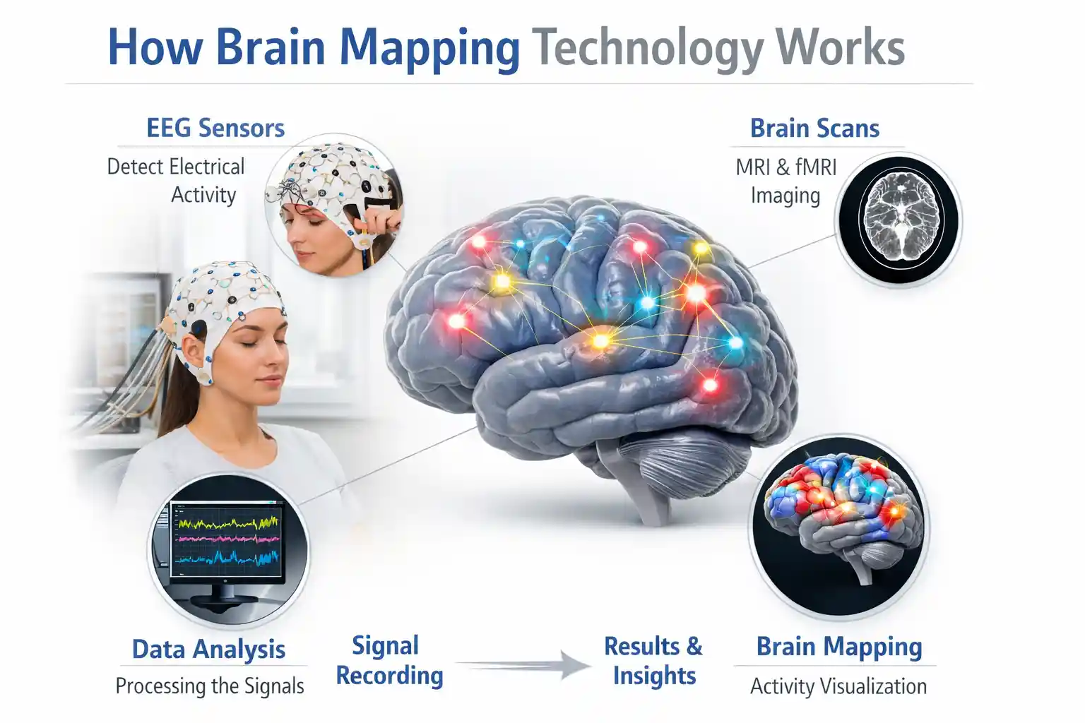
Let’s demystify neuroscience without oversimplifying. Consumer EEG devices measure electrical activity generated by large populations of neurons firing in sync. When you see “alpha waves” or “beta bursts” in an app, you’re seeing aggregated signals from the cortex’s surface—not individual thoughts or emotions.
Research from institutions like Stanford and MIT has shown that certain brainwave patterns correlate with cognitive states. For example, increased alpha power in posterior regions often accompanies relaxed wakefulness, while frontal beta can indicate active concentration. But correlation isn’t causation, and individual variability is huge. A pattern that means “focused” for one person might mean “anxious” for another.
Practical implication: treat device feedback as a directional cue, not a definitive read. If your headset suggests you’re distracted, use it as a prompt to check in with yourself—not as an absolute truth. This nuanced approach aligns with guidance from neuroethics researchers, who emphasize that consumer neurotechnology should augment, not replace, self-awareness.
Current limitations are significant. Consumer EEG can’t reliably distinguish between similar mental states (e.g., focused work vs. stressed rumination) without personalized calibration. Signal quality degrades with movement, sweat, or poor sensor contact. And perhaps most importantly, these devices sample only the brain’s surface—deep structures involved in emotion and memory remain invisible to scalp EEG.
Ethical concerns are emerging, too. Neural data is uniquely intimate. As UNESCO’s global ethics framework on neurotechnology notes, brain data could reveal predispositions, mental health indicators, or cognitive traits that might be misused if not properly protected. While most consumer devices don’t store raw EEG long-term, the potential for privacy erosion is real as the technology advances.
The Honest Drawbacks: What Manuals Don’t Tell You
After weeks of testing, here are the friction points that nearly made me quit:
Physical discomfort: Headbands can cause pressure headaches after 30+ minutes. Earbuds like the IDUN require a snug fit that some users find intrusive. I developed a mild rash behind my ear from the Muse’s sensor pads—a reminder that skin contact isn’t trivial.
Setup fatigue: Even “quick-start” devices require consistent positioning. Miss the sweet spot by a centimeter, and signal quality plummets. This isn’t a one-time calibration; it’s a daily ritual.
Software limitations: Many apps prioritize slick visuals over data transparency. I wanted raw spectral power plots; I got gamified “focus scores” with unclear algorithms. OpenBCI’s software is powerful but demands technical literacy.
Inconsistent readings: Environmental factors matter. A ceiling fan, fluorescent lights, or even a nearby smartphone can introduce noise. I learned to test in the same quiet room at the same time of day for comparable results.
Interpretation anxiety: Seeing a “low focus” score when I felt productive created unnecessary doubt. Without context, metrics can mislead. I eventually stopped checking mid-task and used devices only for pre/post session baselines.
These aren’t flaws in the technology per se—they’re realities of bringing lab-grade tools into uncontrolled environments. Acknowledging them isn’t pessimism; it’s responsible consumer guidance.
Who Should Consider This Technology
Ideal Users: Patient biohackers who enjoy iterative self-experimentation, and dedicated meditation practitioners who benefit from tangible, real-time neurofeedback loops.
Healthcare Applications: As an adjunctive tool for clinician-guided biofeedback therapy (e.g., teaching anxiety patients to recognize and modulate their physiological arousal).
Research Applications: Invaluable for academic prototyping, educational neuroscience, and developing novel Brain-Computer Interface (BCI) algorithms using open-source platforms.
Consumer Use Cases: Guided mindfulness, basic sleep onset optimization, and establishing personal baseline metrics for cognitive routines.
Situations Where Adoption May Not Be Appropriate: Individuals seeking a medical-grade diagnosis for neurological or psychiatric conditions; users with severe skin sensitivities or contact dermatitis from electrode materials; and anyone expecting a “plug-and-play” experience without a willingness to learn basic neurophysiology.
How We Researched This Topic
Research References
• NIH BRAIN Initiative Research
• IEEE Standards Association Wearable Biosensor Framework
• Nature Neuroscience Publications
• Stanford Neural Engineering Research
• MIT Media Lab Neurotechnology Studies
• Frontiers in Human Neuroscience
• University of British Columbia Neuroethics Research
Expert Review Information
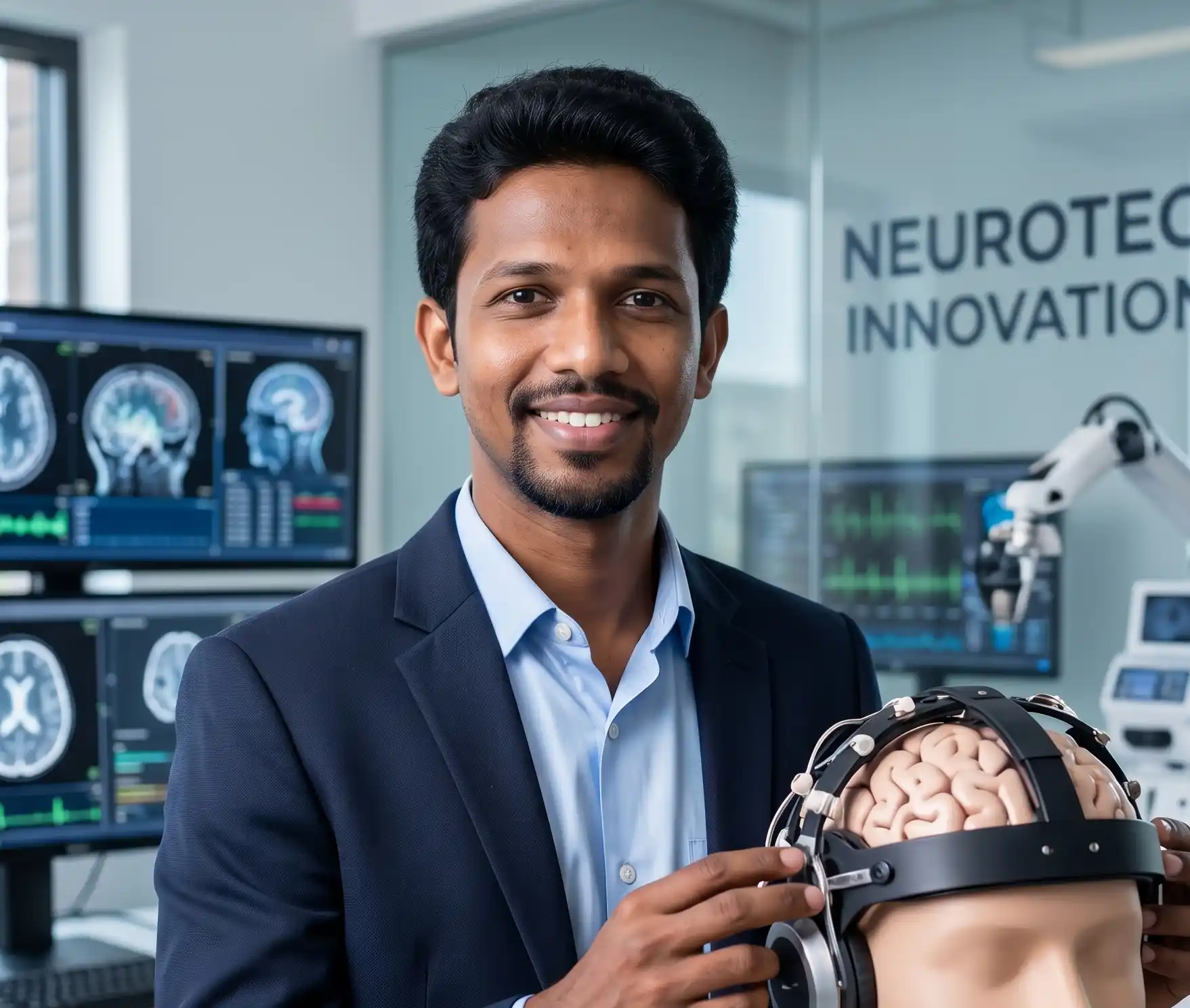
Reviewed By: Asad Ansari
Professional Background: Neurology Technician specializing in EEG, NCV, and neurodiagnostic procedures at Amrita Hospital, Faridabad.
Area of Expertise: Brain Mapping Technologies, EEG Systems, Neurodiagnostics, Neurofeedback, Brain-Computer Interfaces, and Clinical Neurotechnology.
Editorial Review Date: June 2026
Research Sources Evaluated: Clinical studies, neuroscience journals, neurotechnology publications, and evidence-based medical resources.
Disclosure: This content is intended for educational and informational purposes only and should not be considered medical advice, diagnosis, or treatment.
The Bottom Line: Practical Value in 2026
So, is consumer brain mapping technology worth it in 2026? For the right person, with the right expectations, yes, but with caveats.
If you’re curious about your mental patterns and willing to invest time in learning the tools, these devices can offer intriguing insights. They won’t read your thoughts or guarantee peak performance. But they can make abstract states like “focus” or “relaxation” slightly more tangible, creating opportunities for reflection and adjustment.
My recommendation: start small. Try a meditation-focused device like the Muse S if you’re new to neurofeedback. Use it for two weeks without overinterpreting the data. Notice how you feel, not just what the app says. If you find value, consider expanding. If not, you’ve lost little beyond the purchase price.
The field is advancing rapidly. Ear-EEG technology, like the FDA-cleared NAOX LINK, is bringing clinical-grade monitoring into everyday contexts. AI-driven signal processing is improving artifact rejection. But the core challenge remains: translating noisy biological signals into actionable, personalized insights without overselling.
As a researcher who’s tested these tools in the wild, I believe the most valuable outcome isn’t a perfect brain map—it’s a deeper conversation with yourself about attention, rest, and mental well-being. The technology is a mirror, not a map. Use it to reflect, not to dictate.
And if your headset won’t stay put at 6:47 AM? Sometimes, the most human response is to take it off, drink your coffee, and start your day anyway. The brain, after all, was mapping itself long before we built devices to watch.
Important Note
Specific products are referenced as examples of currently available technologies and should not be interpreted as endorsements or purchasing recommendations.